Sulcatas can live over one hundred years, Galápagos tortoises are noted to live over 150 years, but an Aldabra giant tortoise named Adwaita may have been the longest living at an estimated 255 years. In general, most tortoise species can live 80–150 years.
But only a small percentage of these animals live a full life. As with people, turtles and tortoises face a long list of natural diseases.
Recently, we lost the life of one of our loved pets. It is truly heartbreaking. So we wanted to share the following information as well as links to Florida facilities with trained professionals who know how to work on the Chelonian species.
Florida Wild Veterinarian 115 E Euclid Ave, DeLand, FL 32724 (386) 734-9899
Bush Wildlife 2500 Jupiter Park Dr, Jupiter, FL 33458 (561) 575-3399
University of Florida College of Veterinary Medicine 2015 SW 16th Ave, Gainesville, FL 32608 (352) 392-2235
Affiliated Veterinary Specialists 9905 S US Hwy 17 92, Maitland, FL 32751 (407) 644-1287
Respiratory Infections In Tortoises:
Respiratory infections are very common in tortoises. Upper respiratory infections are the most common, but lower respiratory infections (pneumonia) also occur.
One of the most common causes of upper respiratory infections is Mycoplasma bacteria, though other bacteria and viruses may also be causes. Signs include a runny nose, bubbles from the nose, erosion of the nares, swelling around the eyes, and conjunctivitis (reddened, inflamed eyes).
Flushing mucus and exudate out of the tortoise’s sinus cavity along with proper antibiotic therapy is often needed to resolve illness. Tortoises with Mycoplasma are often infected for life, and flare-ups during times of stress or immunocompromise are common.

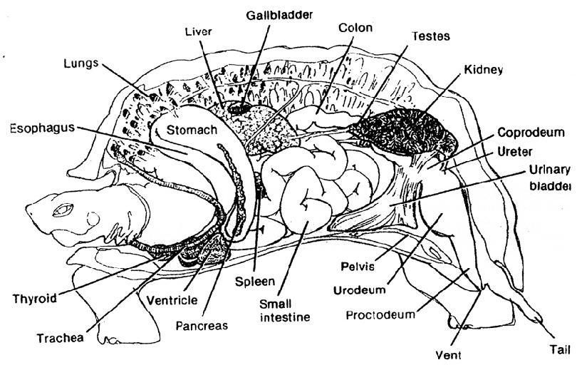
In chelonians, air enters through the nares and passes through the nasal cavities lined by olfactory epithelium and mucosal epithelium.
The glottis is located at the base of the fleshy tongue and often is not visualized in an awake patient. The trachea consists of complete tracheal rings and bifurcates into a left and right unbranched intrapulmonary bronchus at the thoracic inlet.
All chelonians have paired, multichambered lungs that are located underneath the carapace, are relatively rigid, and may extend caudally to the cranial pole of the kidneys. The well-developed bronchi branch into small air passageways that terminate into alveolar tissue.
Many reptiles are presented with chronic respiratory tract disease and by the time of presenting clinical signs include pale, cyanotic membranes and multiple signs of disease may be present including lethargy, anorexia, egg-binding, and dysecdysis. In these patients, additional diagnostic tests are required to fully elucidate the extent of respiratory disease, as well as other organ system function.
Click to see what happens during this vet visit…
Lower respiratory Infections are more significant and require more aggressive treatment. Symptoms include labored breathing (indicated by a tortoise extending its neck and moving its front legs in and out in order to breathe), weakness, lethargy, sunken eyes, and other signs.
Radiographs (X-rays) are usually needed to diagnose lower respiratory infections, and a lung wash to collect samples for cytology and culture should be performed when possible to provide the best treatment.
Traumatic Injuries:
Slow-moving chelonians often fall prey to dogs, other predators, lawnmowers, cars, and intentional injury resulting in cuts, abrasions, cracked shells, broken bones, and internal injuries. Treatment varies with the severity of the injury and time elapsed since the injury. Animals with non-healing wounds are prone to myiasis i.e. infestation with fly maggots. Many of these cases are considered a true emergency. If unable to obtain immediate veterinary care the following first aid measures are indicated:
- Stop any fresh bleeding by applying direct pressure to the wound
- Clean wounds using sterile saline solution (saline solutions used for contact lens work well) and dilute Betadine solution
- If maggots are present, manual removal of the maggots followed with flushing the wounds with hydrogen peroxide first then with sterile saline solution and dilute betadine
- Silvadene (1% Silver Sulfadiazine Creme) or another topical antibiotic ointment should be applied to the wounds
- Cover with telfa pads and apply pressure type wrap using masking tape or cellophane wrap to prevent oozing and further contamination
- Seek veterinary attention as soon as possible- Injectable or oral antibiotics and surgical repairs are often indicated.
Infectious Disease:
Signs of difficult respiration include stretching the neck, gaping the mouth with labored breathing and pumping their head and legs while breathing. Aquatic turtles may float unevenly with one side floating higher than the other in the water. Nasal discharge is abnormal unless associated with drinking. Bacterial infections are a common cause of infections of the upper respiratory tract and can result in death if not treated early on in the course of the disease. Poor husbandry, suboptimal environmental conditions, and Vitamin A deficiency can predispose to respiratory infections.
Vitamin A deficiency is common in reptiles, especially water turtles on a diet of dried insects and lettuce. Vitamin A is important in the survival and function of the cells that line the respiratory tract, digestive tract, mucous membranes and glands of the eyes.
Vitamin A deficiency may present as swollen eyes, increased mucus in the mouth and flaky skin. These animals are predisposed to secondary bacterial infections leading to mouth rot, shell rot and pneumonia. Foods high in Vitamin A include cooked liver, cod liver oil, yellow vegetables, carrots, egg yolk, and Brussels sprouts.
Nutritional Deficiencies:
Vitamin B1 (Thiamin) – Tremors, paralysis, other neurologic signs. Frozen fish contains thiaminase, an enzyme that destroys thiamin.
Vitamin C– Increased incidence of infection.
Vitamin K– Bleeding problems.
Vitamin D– Low Vitamin D causes a lack of absorption of calcium in the intestine. High Vitamin D causes calcification of soft tissues leading to organ failure.
Protein– Too little causes poor growth and poor immune function i.e. low disease resistance. Too much contributes to early kidney disease.
Disorders of the Digestive Tract:
Failure to pass feces may be due to dehydration; impaction with foreign material such as sand, gravel, or cage litter; or starvation. Diarrhea may result from feeding foods with high water content, parasites, overgrowth of abnormal bacterial organisms and lack of adequate roughage in the diet of tortoises.
Gas may accumulate in the bowel due to an abrupt dietary change, inappropriate foods (dairy products), low body temperature or secondary to gastrointestinal obstruction resulting in bloat. Infections of the mouth (stomatitis) are usually due to bacterial infections, however, these are relatively uncommon in chelonians. Masses protruding from the rectum (anus) should be considered an emergency. This can be associated with a prolapse of the rectum, bladder, hemipenes (penis) in male chelonians, and uterus in females.
Reproductive Disorders:
Egg binding or dystocia, the inability to deliver eggs, is a common problem in captive chelonians. The causes include the absence of normal substrate for the turtle to nest in, abnormal shape or size of the egg, soft eggs, broken eggs, disease of the uterus causing scaring or other obstruction, and blockage of the pelvis by bladder stones, intestinal impaction or foreign body preventing normal delivery.
The most common signs observed are loss of appetite and straining. Treatment varies with the cause of the dystocia from the provision of proper nesting substrate to surgical removal of eggs. Failure to relieve dystocia can result in death. Straining as a result of eggs, intestinal foreign bodies or bladder stones may result in prolapse of the rectum, bladder or uterus. Treatment involves cleaning the prolapsed tissue with sterile saline solution and coating the tissue with a water-soluble lubricant such as K-Y jelly to prevent drying and trauma.
Granular sugar may be used to draw fluid out of the edematous tissues. Keep the tissues moist and present the animal to a veterinarian as soon as possible.
The shorter the time that the tissue is exposed the better the chance that the tissue will survive and be successfully replaced. Male chelonians often prolapse their penis as a part of courtship display. In these cases, the penis usually returns to its normal position unaided, however, if drying or injury occurs, assistance may be needed.
Metabolic Bone Disease:
Metabolic Bone Disease (MBD) is the loss or lack of normal bone density resulting from one or a combination of the following factors: low dietary calcium, high dietary phosphorus, low Vitamin D3, and/or kidney disease. Vitamin D3 is required for normal absorption of calcium from the digestive tract. Low Vitamin D3 is caused by lack of exposure to natural sunlight.
The body constantly tries to balance calcium with phosphorus in the blood in order to provide appropriate calcium for vital cellular functions. If calcium is not readily available in the diet, then the body pulls calcium from the bones.
In growing animals, this results in deformity of bones and shell, stunting and if severe, death. In adults, the bones are brittle and easily broken, the shell may become soft, intestinal motility may be reduced and egg binding is more common.
Prevention by feeding a proper diet is best. Foods with a normal calcium balance (2 parts calcium to 1 part phosphorus) include zucchini, fig, endive, alfalfa hay/pellets, pinkie mice, earthworms, mealworms, crickets, and slugs. Many zoos feed rabbit or guinea pig pellets. These alfalfa pellets are good sources of calcium, protein and fiber/roughage.
Parasites In Tortoises:
Parasitic health-related problems are relatively common in tortoises, and parasites can infect the intestinal tract, tissues of the body, or feed on the shell and skin. Not all parasites cause harm to tortoises; some may even be beneficial to certain tortoises.
Diagnosing parasites in tortoises can require tests different from those performed frequently on mammals. With regard to intestinal parasites, I highly recommend testing at least one fecal sample, and a second sample one month later, for parasites after acquiring a new tortoise, and especially before placing it in an enclosure with other existing tortoises.
The fecal sample being tested should be recently passed, kept moist by wrapping it in a wet paper towel placed in a plastic bag and not allowed to get too hot or cold. Time outside of the body, drying, heat, and cold can decrease our ability to detect certain types of intestinal parasites. Make sure your veterinarian performs a “direct” fecal test looking for protozoans in addition to the standard floatation test.
Different types of parasites require different types of treatments, so it is important to identify and treat parasites of concern properly. Routine deworming is becoming less popular due to increasing parasite resistance to medications, the understanding being that not all parasites are problematic and administration of anti-parasitic medications can potentially cause illness if a tortoise is not dosed properly or if the animal has a reaction to them.
Certain types or low numbers of intestinal protozoans are often normal, do not cause problems, and do not require treatment. Pinworms are present in most tortoises and rarely need treatment. These should be differentiated from other nematode parasites in which treatment is often recommended, however.
Also, numbers of “normal” parasites may increase to levels that cause illness when tortoises are not provided proper diets and husbandry. Many tortoises, for instance, are fed too much produce and need more grass or high-fiber plant leaves and stems in their diet.
Feeding, drinking, and defecating in the same small area day after day can lead to a parasite load that’s higher than normal. Tortoises often like to sit in their water bowls, and defecate and drink at the same time.
It’s important that you work with a knowledgeable reptile veterinarian, and it is also important that routine cage cleaning and sanitation occurs.
Generalized treatments include:
- Betadine soaks- Dilute betadine to light tea color and use as a soak solution for 20 minutes twice a day
- Silvadine (1% Silver Sulfadiazine Creme)- Apply to affected areas twice a day
- Keep dry other than during feeding (water turtles only) and between betadine soak
- Soak in fresh water twice a day to encourage drinking, defecation, and urination in most species and to feed aquatic turtles
- Injectable or oral antibiotics may be prescribed by the veterinarian based on observation, clinical signs and bacterial/fungal culture and sensitivity testing results when possible
- The fungal disease may respond to saltwater soaks (1/4 cup non-iodized salt to 5 gallons of water) and to dilute nolvasan soaks
Septicemia:
Septicemia is often undiagnosed in reptiles, resulting in improper and unsuccessful treatment of the patient. In reptiles, septicemia most commonly originates from the gastrointestinal, urinary, or respiratory tracts.
A presumptive diagnosis of septicemia can be made by physical findings, but a venous blood sample, aseptically collected and submitted for culture and sensitivity testing, is needed for confirmation. In some cases, it is indicated to treat the animal for suspected septicemia while laboratory results are pending.
Broad-spectrum antimicrobial agents effective against common reptilian pathogens should be selected. It may become necessary to adjust antimicrobial drug selection based on culture and sensitivity results.
Leave a Reply